Take care of your breast health at the Breast Imaging Center
Each year, hundreds of thousands of Americans – both men and women – are diagnosed with breast cancer. It can be scary to be screened for breast cancer. Let our breast care specialists walk you through the process and support your journey to getting answers.
At the Breast Imaging Center at NMC Health’s Medical Center, our team of expert technologists offer a variety of diagnostic testing options for breast care. Services include:
- Digital 3D mammography-Digital Breast Tomosynthesis
- Bone Density Scan (DEXA)
- Ultrasound/Sonography
- Breast MRI
- Stereotactic-guided breast biopsies
- Ultrasound-guided breast biopsies
- Wire localizations
If you have a medical emergency, call 911 or visit the NMC Health Medical Center Emergency Department.
When scheduling your mammogram
When you schedule your exam we will need to know if you have any concerns about your breasts. Are you scheduling for your annual mammogram? If you have any concerns- any “new” pain, lumps or discharge, you should consult your provider prior to scheduling. There are different kind of mammograms and we want to be sure that you get the correct exam. Also, it is important to let us know if you have implants. If you do, we need to give you some special considerations and want to ensure that you receive the optimal experience.
Allow about thirty minutes to an hour for your full appointment.
Small children will not be allowed in the exam room, so we ask you find someone to care for them on the day of your exam. If you cannot find child care, we may need to reschedule your appointment.
If breast tenderness is a problem for you, consider avoiding caffeine for a week before your mammogram.
We will ask you to remove your deodorant for the exam. It will show up in your images. Please feel free to wear it to your appointment, we will have wipes available for you to wipe it off.
Diagnostic Breast Imaging
When visiting the NMC Health Breast Imaging Center, you have a variety of options available to you for diagnostic breast imaging. Click on the types below to see a description of what each are used for to detect breast issues.
NMC Health added 3D mammography to its breast imaging services in 2015. 3D mammography or digital mammography is an advanced, clinically proven screening and diagnostic tool designed for early breast cancer detection.
During the 3D mammography exam, the X-ray arm sweeps in an arc over your breast, taking multiple low-dose images. Then, a computer produces a 3D image of your breast tissue in one millimeter slices, providing greater visibility for your radiologist to see breast detail in a way never before possible. This significantly improves early breast cancer detection and lowers the need for follow-up imaging by up to 40%, which can reduce unnecessary anxiety.
Ultrasound imaging of the breast uses sound waves to produce pictures of the internal structures of the breast. The primary use of breast ultrasound is to help diagnose breast abnormalities detected by a physician during a physical exam (such as a lump) and to characterize potential abnormalities seen on mammography or breast magnetic resonance imaging (MRI).
Ultrasound imaging can help to determine if an abnormality is solid, fluid-filled (such as a benign cyst) or both cystic and solid.
An MRI of the breasts uses a strong magnetic field and radio frequency waves to produce detailed pictures.
It is primarily used for patients at high risk for breast cancer, evaluating breast implants for rupture or to further evaluate abnormalities seen on mammography.
MRI of the breast is not used in place of mammography or ultrasound imaging but is used as an additional tool to further evaluate certain breast conditions.
A breast biopsy is a simple procedure to remove a small sample of breast tissue for laboratory testing. The type of biopsy recommended depends on a number of things, such as:
- Can the abnormality be best seen with mammography, ultrasound/sonography or MRI?
- How suspicious the breast change looks
- How big it is
- Where it is in the breast
- If there is more than one
- Any other medical problems you might have
After your biopsy, a physician will use your tissue sample to identify and diagnose abnormal cells that make up breast lumps or other concerning findings on a mammogram, ultrasound or MRI. Your breast biopsy results can help determine whether you may need additional surgery or other treatment.
NMC Health Breast Imaging Center performs two types of breast biopsy procedures. Click on the types below to see a brief description of each.
Lying face down on a table, your breast will be compressed to take pictures to locate the abnormality. After the biopsy site is clean, the physician will numb the area.
A special needle will then be inserted into your breast using digital imaging to guide the placement of the needle, thus obtaining multiple small tissue samples.
Lying on your back, your breast will be imaged to locate the abnormality. After the biopsy site is clean, the physician will numb the area.
A special needle will then be inserted into your breast using ultrasound to guide the placement of the needle, thus obtaining multiple small tissue samples.
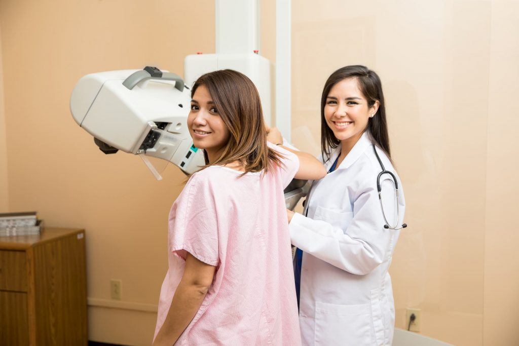
What to expect during a mammogram
You might be getting ready to have your first mammogram, which can cause some feelings of anxiety. Here’s what to expect during a mammogram, so you are prepared.
When you arrive at NMC Health Medical Center, park in front of the hospital and enter at the hospital’s main entrance. Check in at Registration where you will be asked to provide your photo ID and insurance information. Please arrive 30 minutes prior to your appointment time to ensure this process runs smoothly.
The technologist will talk you through the entire exam and tell you what he or she will be doing during the procedure. You will be positioned on to the mammogram machine. The machine will put compression on your breasts for a brief period of time while it takes pictures of the soft tissue. This should not be painful, but it might be slightly uncomfortable. It’s important to hold still so the images come out crisp and clear. You might be asked to hold your breath during the mammogram.
If you’re feeling any pain or discomfort during your exam, let your technologist know. Some people with back, neck or shoulder problems might be uncomfortable, so tell your technologist before you begin so they can accommodate your needs.
You can expect a letter in the mail regarding the results of your exam within three to seven days. If the radiologist sees any changes in your breast since your last mammogram, or finds an abnormality, we will contact you to schedule additional studies.
If you have any questions, please feel free to speak with our imaging staff.
Mammography Accreditation
The ACR gold seal of accreditation represents the highest level of image quality and patient safety. It is awarded only to facilities meeting ACR Practice Guidelines and Technical Standards after a peer-review evaluation by board-certified physicians and medical physicists who are experts in the field.
FAQs
When it comes to breast health, getting a yearly mammogram can help detect breast cancer or breast changes early. Every year, hundreds of thousands of people are diagnosed with breast cancer. It’s the second leading cause of cancer death in women. One in eight women and about one in 800 men will be diagnosed with breast cancer at some point in their life.
Getting routine mammograms and breast screenings can reduce your risk of death if you should develop breast cancer. Early detection is crucial in being able to treat the cancer before it grows and spreads.
The National Cancer Society recommends you start getting annual mammograms and breast screenings at the age of 40.
3D mammography is the most advance technology available in breast care. Using high-resolution images, a readiologist can detect and pinpoint any breast abnormalities at a highly accurate rate. This technology uses X-ray technology to look into your breast tissue. The machine is the same as what you are used to, but the images are clearer, giving your radiologist a better picture to work with.
The amount of radiation used during a routine mammogram is small. If you have concerns about the radiation or have a special condition, please make sure your radiology technologist knows about it before you have your mammogram. He or she will be able to answer any questions and address any concerns.
In most cases, your mammogram will be covered under your insurance as preventative care. Check with your insurance company to see if you can expect to pay anything out-of-pocket for the procedure.
Medicare covers routine mammograms for women over the age of 40 without a co-pay. If you struggle with low income, the National Breast and Cervical Cancer Early Detection Program may cover the cost of your mammogram. Contact the Harvey County Health Department for more information.
Breast Imaging Library

5 tests that can save your life

5 ways to lower your risk of breast cancer

A Message from the CEO: Breast Cancer Awareness
NMC Health Earns Radiology Accreditation
3D Mammography Now Available at Newton Medical Center